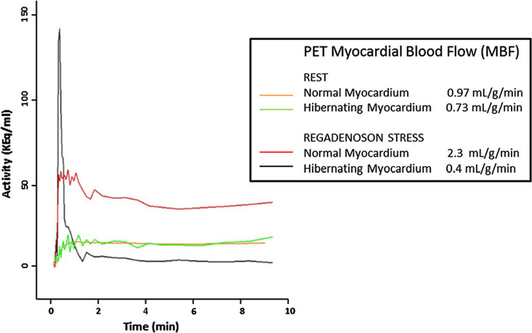
Figure 3.
Quantification of myocardial blood flow (MBF) by PET. Rest and stress MBF was quantified in mL/g/minute using the 13N-ammonia PET data sets. At rest, MBF in normal and hibernating myocardium were comparable (0.97 vs 0.73 mL/g/minute). With stress MBF in hibernating myocardium was paradoxically reduced compared to resting blood flow (from 0.73 to 0.4 mL/g/minute), indicative of a coronary artery steal phenomenon.

