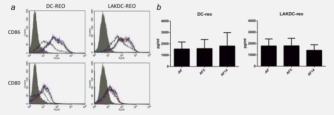
Figure 5.

LAKDC and reovirus phenotypically mature iDC and produce proinflammatory cytokines in the presence of ascites. (a) Reovirus-loaded iDC or LAKDC were cultured in the absence (red line) or presence of 2.5% AF5 (green line) or AF14 (blue line) for 48 hr. DC (CD11c+ cells) were analyzed by flow cytometry for expression of maturation/activation markers, CD80 and CD86. Histogram plots are representative of two healthy donors. Shaded gray = isotype control, black line = unloaded DC controls [either iDC (left panel) or DC within LAKDC coculture (right panel)]. (b) Reovirus-loaded iDC and LAKDC were cultured for 48 hr ± ascites (AF5 and 14) before supernatants were collected and concentrations of IFNα were determined by ELISA. Graphs show the mean + SEM of four healthy donors.
