
Figure 4.
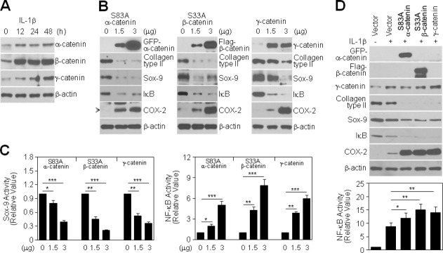
Role of IL-1β-induced catenin proteins in chondrocyte destruction. (A): Chondrocytes were treated with 10 ng/mL IL-1β for the indicated periods. Levels of catenin proteins were detected by Western blotting. (B, C): Chondrocytes were transiently transfected with GFP-tagged S83A α-catenin, FLAG-tagged S33A β-catenin, or wild-type γ-catenin, as indicated, for 48 h without (B) or with a Sox-9 (C, left) or NF-κB reporter gene (C, right). Levels of differentiation- and inflammation-associated proteins were determined by Western blotting (B), and Sox-9 or NF-κB transcriptional activity was determined by reporter gene assay. Data are expressed as means ± SDs (*P < 0.05, **P < 0.005, ***P < 0.0005 compared with untransfected controls) (C). (D): Chondrocytes were transiently transfected with 3 μg of each catenin plasmid, as indicated, for 24 h and then left untreated (−) or treated (+) with 10 ng/mL IL-1β for an additional 48 h. Levels of differentiation- and inflammation-associated proteins were determined by Western blotting (top), and NF-κB transcriptional activity 24 h after IL-1β treatment was determined by reporter gene assay. Data are expressed as means ± SDs (*P < 0.05 and **P < 0.005 compared with cells treated with IL-1β alone) (bottom).