
Figure 1.
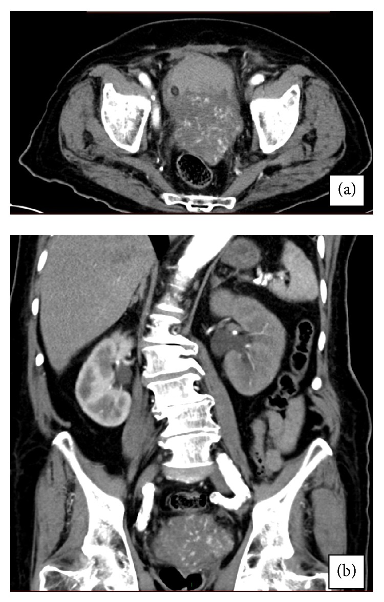
Computed tomography of our patient. (a) Highly vascular mass originating from the left bladder wall, but a prostatic origin could not be absolutely rolled out by CT. (b) Hydronephrosis on the left side and coronal view of the mass.
Official websites use .gov
A
.gov website belongs to an official
government organization in the United States.
Secure .gov websites use HTTPS
A lock (
) or https:// means you've safely
connected to the .gov website. Share sensitive
information only on official, secure websites.
Computed tomography of our patient. (a) Highly vascular mass originating from the left bladder wall, but a prostatic origin could not be absolutely rolled out by CT. (b) Hydronephrosis on the left side and coronal view of the mass.