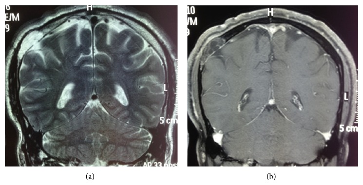
Figure 2.

(a) T2 MRI showing high signal within the intraosseous lipomatous meningioma and dural defect. (b) T1 MRI with gadolinium observing heterogenous uptake within the lipomatous meningioma with no obvious intraparenchymal extension.

(a) T2 MRI showing high signal within the intraosseous lipomatous meningioma and dural defect. (b) T1 MRI with gadolinium observing heterogenous uptake within the lipomatous meningioma with no obvious intraparenchymal extension.