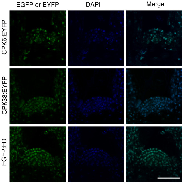
Figure 5. Spatial expression patterns and subcellular localization of FD, CPK6, and CPK33 in shoot apical meristem.

Shoot apical meristem in 7-day-old plants of pCPK6::CPK6:EYFP:3HA:His, pCPK33::CPK33:EYFP:3HA:His, and pFD::EGFP:FD; fd-1 are shown. EYFP (CPK:EYFP) or EGFP (EGFP:FD) fluorescence, DAPI fluorescence, and merged images are shown in the same magnification. Scale bar: 50 μm.
