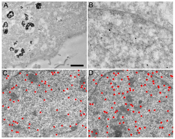
Figure 1. Transmission electron micrographs of HeLa cell sections labeled in vivo with antibodies directed against RNA polymerase II and coupled to gold beads.

(A) Antibodies coupled to 6 nm colloidal gold particles are found aggregated in large cytoplasmic vesicles. (B) Antibodies coupled to 0.8 nm gold particles are detected as individual spots after silver enhancement in the cytoplasm and are enriched in the nucleus. (C) Distribution of the amplified gold particles coupled to IgG molecules directed against RNA polymerase II. (D) Distribution of the amplified gold particles coupled to Fab fragments directed against RNA polymerase II. The bar represents 1 μm in (A), 0.2 μm in (B) and 0.6 μm in (C) and (D).
