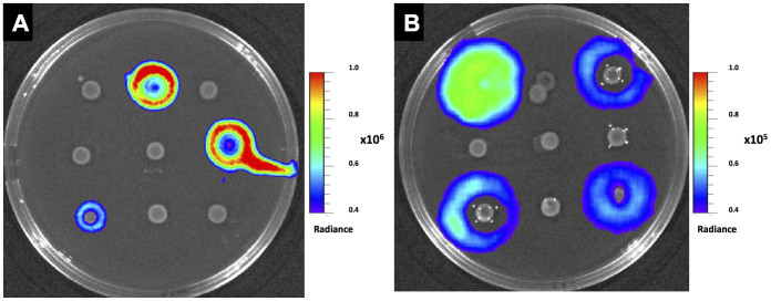
Figure 2. Bioluminescence-based detection of CylLS (A) and GBAP (B) producers.
From the right to the left, uppermost row: V583, DS5, and E1Sol; central row: V583fsrB*, CH188, and ×98; bottom row: MHH594, T2, and V583ΔgelE. The E. faecalis isolates were cultured on GM17 agar plates, and the biosensor was overlaid after an overnight incubation. Plates were kept at 37°C for 3 hours and imaged with a Xenogen IVIS Lumina II Imaging System (Calipers Corp., CA). A 10-fold higher light emission was induced by CylLS than by GBAP.

