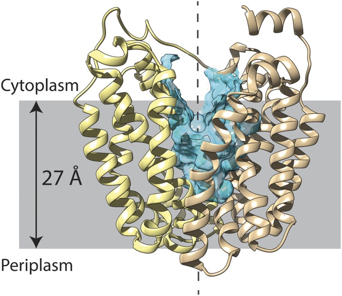
Fig. 1.
LacY ribbon presentation in an inward-open conformation with a twofold axis of symmetry (broken line). (Left) N-terminal helix bundle (light yellow). (Right) C-terminal helix bundle (tan). The cytoplasmic side is shown at the top. The blue region represents the hydrophilic cavity, and the gray-shaded area represents the membrane.

