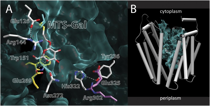
Fig. 6.
Crystal structure of single-Cys122 LacY with covalently bound MTS-Gal. (A) Side chains are shown as sticks. The yellow side chains (Glu269 and Trp151) make direct contact with the galactopyranosyl ring of MTS-Gal covalently bound to a Cys at position 122. The gray side chains are not sufficiently close to make contact with the galactopyranosyl ring. Glu325 and Arg302 (in purple) are involved in H+ transport. The green felt-like area represents the Van der Waals lining of the cavity. Note that the periplasmic side is closed. (B) Structure of single-Cys122 LacY with covalently bound MTS-Gal viewed from the side. Helices are depicted as rods, and MTS-Gal is shown as spheres colored by atom type with carbon in green. The aqueous central cavity open to the cytoplasmic side is colored light green.

