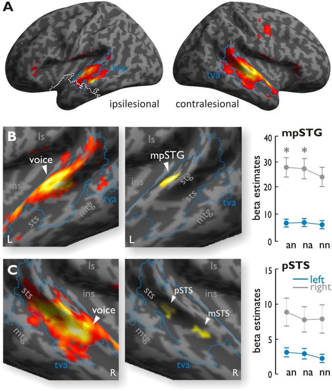
Fig. 2.
(A) Activity for vocal sounds compared with nonvocal sounds across all MTL patients in experiment 1. The blue outline denotes the temporal voice area (tva), and the white line denotes the lesion extent. (B, Left) In right MTL patients, comparing voices relative to nonvocal sounds showed extended activity in the left STG. (B, Middle) When comparing right MTL patients relative to left MTL patients, voices produced significantly higher activity in the left mpSTG. (B, Right) Activity in the functionally defined voice-selective STG, plotted across experimental conditions during the dichotic listening task (experiment 2), also showed increased activity for an and na trials compared with nn trials for the right MTL patients (as indicated by the asterisks) but not for the left MTL patients. (C, Left) In left MTL patients, comparing voices relative to nonvocal sounds showed extended activity in the right STG. (C, Middle) Comparing responses to voices in left MTL patients relative to right MTL patients activated the right pSTS and mSTS, although with a slightly lower voxel threshold of P < 0.006 and cluster extent of k = 77. These clusters were not modulated by the experimental conditions during the dichotic listening task (experiment 2). ins, insula; L, left; ls, lateral sulcus; mtg, middle temporal gyrus; R, right.

