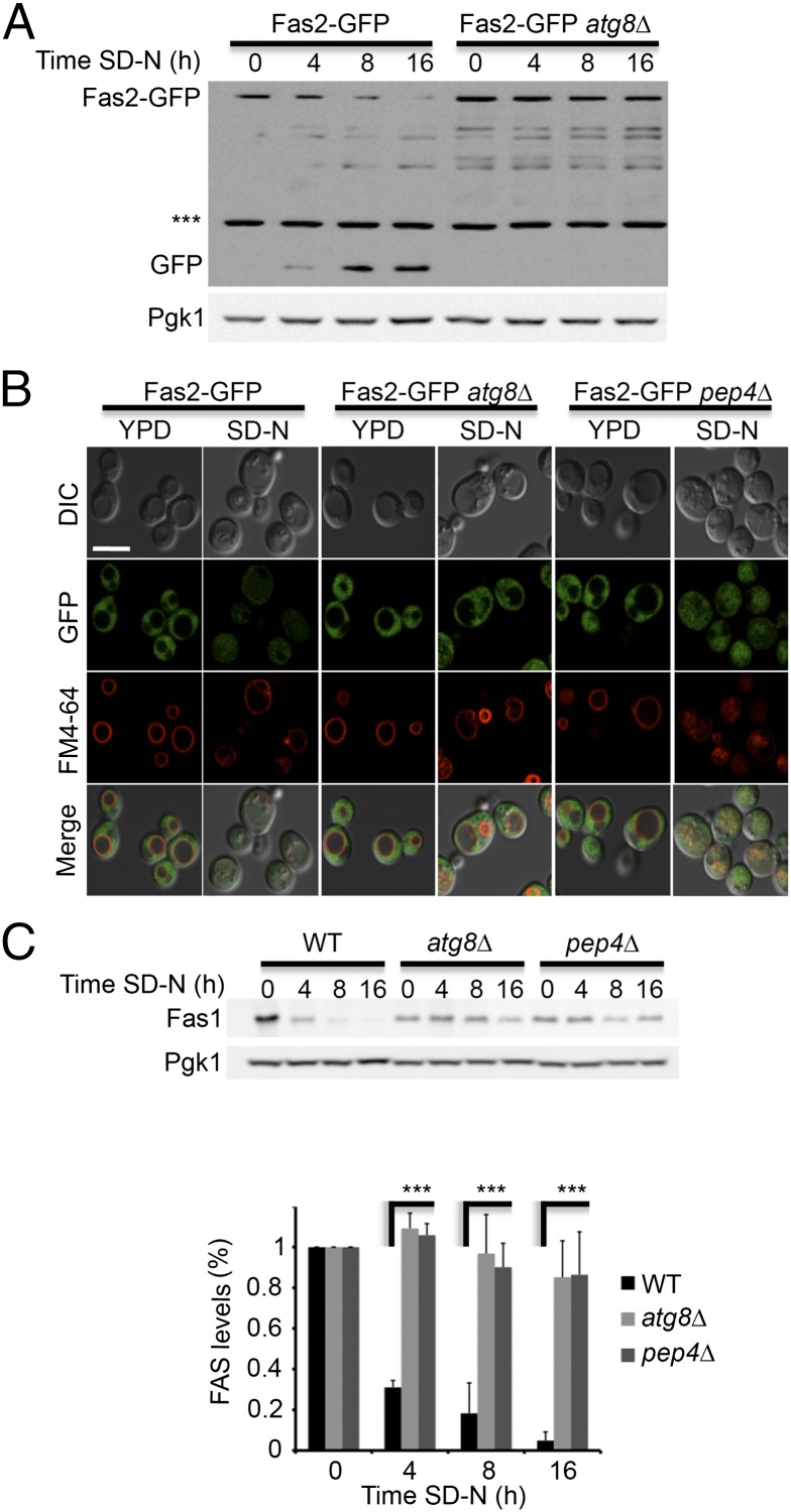
Fig. 2.
FAS degradation depends on the core autophagy genes. (A) FAS2-GFP (TOS017) and FAS2-GFP atg8∆ (TOS023) S. cerevisiae cells were grown to midlog phase and shifted to SD-N medium for the indicated time periods. Cell lysates were subjected to SDS/PAGE, followed by Western blot analysis using anti-GFP and anti-Pgk1 antibodies. (B) FAS2-GFP (TOS017), FAS2-GFP atg8∆ (TOS023), and FAS2-GFP pep4∆ (TOS022) cells were grown to midlog phase and shifted to SD-N medium. Fas2-GFP localization was monitored in YPD medium and after 16 h in SD-N. FM4-64 was used to detect vacuolar membranes. (Scale bar, 5 µm.) (C) WT (BY4741), atg8∆ (TOS006), and pep4∆ (TOS015) cells were grown to midlog phase and shifted to SD-N medium for the indicated time periods. Cell lysates were subjected to SDS/PAGE, followed by Western blot analysis using anti-Fas1 and anti-Pgk1 antibodies. Error bars represent the SDs of three independent experiments. ***P < 0.001 (Student’s t test) (Lower).

