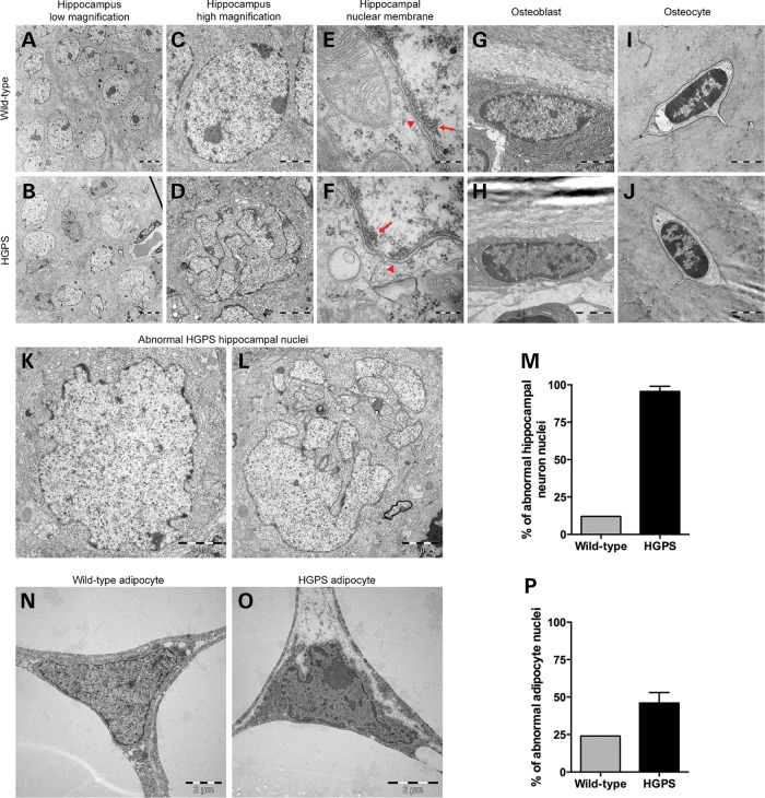
Figure 4.
Electron microscopic analyses of nuclei of various cell types from 70-week HGPS and wild-type animals. While the wild-type hippocampus nuclei from the CA1 region consistently had a smooth round shape (A and C), the HGPS hippocampal nuclei of the CA1 region had a very irregular shape (B and K) and presented with extensive folding, blebbing and lobulation of the nuclear envelope, which was so severe that the nuclei looked fragmented (D and L). The images from the nuclear membranes (arrowheads; E and F) of the hippocampal neurons in the wild-type (E) and HGPS (F) animals showed no apparent loss of heterochromatin (arrows). (G–J) There were no significant differences in the nuclear structure of osteoblasts or osteocytes. (M) Quantification of abnormal nuclei in the hippocampus showed that 95.5% of hippocampal neurons had abnormal nuclei, compared with 11% in wild-type animals. (N and O) Nuclei of adipocytes from HGPS animals showed moderate folding and irregularity in nuclear morphology compared with wild-type animals. (P) Quantification of the percentage of adipocytes with abnormal nuclei morphology.

