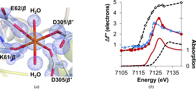
Figure 5.
Characterization of the MMB site. (a) The 2F o − F c electron-density map (light blue) at the MMB site, with Fe16 and coordinating ligands contoured at 3σ. The C atoms are highlighted in gray, N atoms in blue, O atoms in red and Fe atoms in orange. (b) Top: comparison of the refined Δf′′ spectrum of Fe16 (red solid line with red circles) with that of the averaged Fe in the P cluster (black dashed line with white diamonds) of Cp1 and Fe16 in Av1 (blue dashed line with blue triangles). Bottom: the XAS spectra of ferrous sulfate heptahydrate (FeSO4.7H2O, red solid line) and ferric sulfate hydrate [Fe2(SO4)3.xH2O, black broken line] (Zhang et al., 2013 ▶). No background removal or normalization were applied to the refined Δf′′ spectra.

