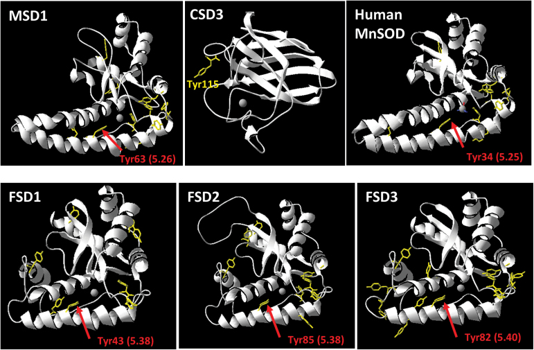
Fig. 9.
Structural model of MSD1, CSD3, human MnSOD, FSD1, FSD2, and FSD3. The structural model of Arabidopsis SODs was generated using SWISS-MODEL with the crystal structure of Caenorhabditis elegans MnSOD as template (PDBcode: PDB 3DC6). The active site ion is shown in grey. All Tyr residues are highlighted in yellow. Tyr63 of MSD1 and the corresponding tyrosine residues in FSD1 (Tyr43), FSD2 (Tyr85), FSD3 (Tyr82), and human MnSOD (Tyr34) are marked with a red arrow. The distance to the active site ion is given in brackets. Tyr115 of CSD3 is indicated in yellow.

