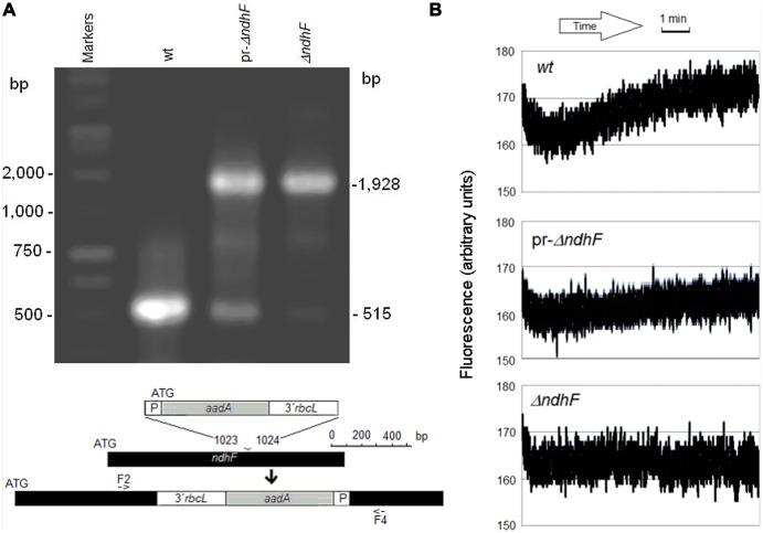
FIGURE 1.
Genetic identification and fluorescence properties of pr-ΔndhF tobacco. (A) PCR amplification products of plastid DNAs of wt, pr-ΔndhF, and ΔndhF tobacco plants using primers F2/F4 (Serrot et al., 2012) for the ndhF gene sequence (bottom map). Sizes of the main amplified fragments and of some markers are indicated on the left and right, respectively. (B) Chlorophyll fluorescence traces of wt, pr-ΔndhF, and ΔndhF tobacco plants after relative high to minimum light transition. Assays were performed with intact tobacco leaves as described Martín et al. (2009). The traces shown are of fluorescence readings every 0.1 s during the final 9 min of minimum light. Vertical axes show the relative fluorescence readings.

