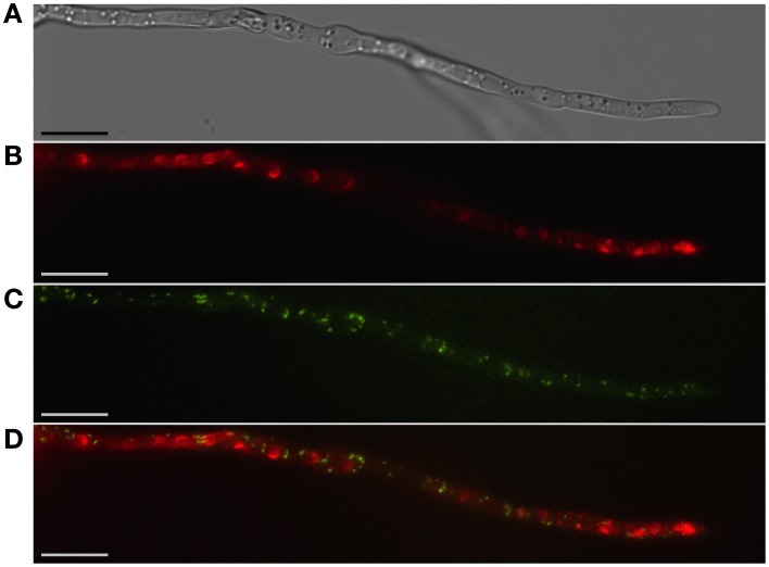
Figure 2.
Localization of peroxisomes and toxisomes in F. graminearum. Shown is a strain of F. graminearum having a GFP-tagged Pex3 protein and a TagRFP-T-tagged trichodiene oxygenase grown under trichothecene-inducing conditions. (A) Hypha visualized using differential interference contrast (DIC) microscopy. (B) TagRFP-T visualized by epifluorescence reveals the trichodiene oxygenase in spherical toxisomes in the subapical cells and in reticulate pattern toward the hyphal tip. (C) GFP fluorescence from Pex3 revealing puntate structures corresponding to peroxisomes. (D) Overlay of GFP and TagRFP-T fluorescence showing that peroxisomes are distinct from toxisomes. Bar = 10 μm. Results presented in Menke et al. (2013); figure generated for Weber (2013).

