Figure 5.
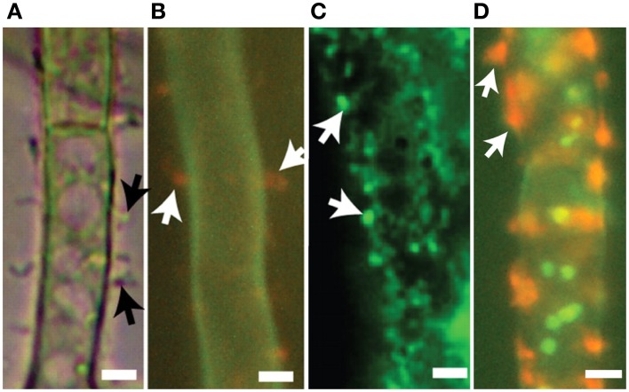
Export vesicles and localized secretion of aflatoxin and norsolorinic acid. Toxigenic cells of A. parasiticus. (A) Visualized using bright-field microscopy, toxigenic cells show pronounced extracellular protuberances (arrows). (B) Similar toxigenic cells treated with the fluorescent vital dye FUN-1 visualizing orange extracellular bodies (arrows) normally associated with cylindrical intravacuolar structures (Millard et al., 1997). (C) Detection of foci (arrows) on surfaces of toxigenic cell by immunofluorescence using anti-aflatoxin antibodies. (D) Detection of norsolorinic acid (NA) on the cell surface (arrows) using fluorescent anti-NA antibodies. Photos reprinted from Chanda et al. (2010) by permission from the publisher, the American Society for Microbiology.

