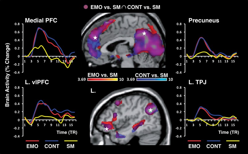
Fig. 3.
The AM retrieval network. Compared with SM, retrieval of AMs, with either Emotion (EMO) or Context (CONT) focus, yielded overlapping increased activity in typical regions of the AM retrieval network. As also illustrated in Table 2, this network included midline cortical structures (medial PFC and precuneus), lateral frontal, temporal and parietal areas, as well as MTL structures (amygdala and hippocampus, data not shown). The activations maps for Emotion vs SM and Context vs SM were set up at a threshold of P < 0.001/t ≥ 3.69 (k ≥ 10 voxels), and superimposed on high-resolution brain images displayed in sagittal views. The coloured horizontal bars show the gradient of the t-values. The line graphs show the time course of responses from typical AM retrieval areas, as extracted from peak voxels (indicated by the white stars) reaching a FWE-corrected threshold of P < 0.05 (t ≥ 6.78) for the AM vs SM contrast (see Table 2, for all peak locations). PFC, prefrontal cortex; vlPFC, ventro-lateral PFC; TPJ, temporo-parietal junction; L, left; TR, repetition time (1 TR = 2 s).

