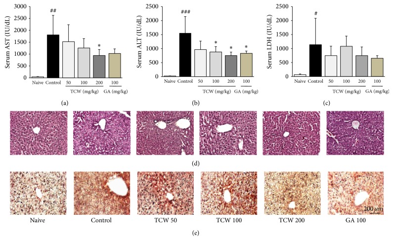
Figure 2.
Serum biochemistries and histopathological and immunohistochemical findings of hepatic tissue. The mice were administered with TCW (50, 100, and 200 mg/kg), gallic acid, or distilled water orally once per day for 5 days. After 18 h of t-BHP injection, serum AST (a), ALT (b), and LDH (c) levels were measured. H&E staining (d) and anti-4-HNE staining (e) were conducted and examined under microscopy (×200). Data are expressed as the mean ± SD (n = 6). # P < 0.05, ## P < 0.01, and ### P < 0.001 compared with the naive group; * P < 0.05 compared with the control group.

