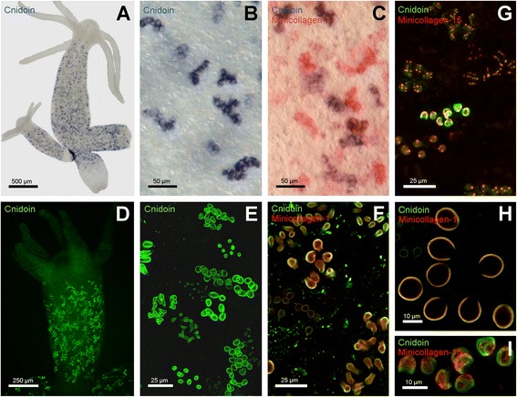
Figure 2.

Gene expression pattern and immunohistochemical localisation of Cnidoin. (A) gene expression pattern as shown by in situ hybridisation. The signal is detected in nests of developing nematocytes in the body column. (B) close-up of nematocyte nests shown in A. (C) double in situ hybridisation of Minicollagen-1 (red) and Cnidoin (blue). (D) Antibody staining reveals nests of developing nematocytes in the body column of Hydra. (E) enlarged view of the immunostaining shown in D. (F) costaining of Minicollagen-1 and Cnidoin in PFA-fixed animals. Both signals localise to the capsule wall. (G) co-staining of Minicollagen-15 and Cnidoin in Lavdovsky-fixated animals. This fixation reveals the presence of both proteins in the tubule of developing nematocysts. (H) enlarged view of a nest with Cnidoin and Minicollagen-1 immunostaining. (I) magnification of a nest with a tubule-specific signal for Cnidoin and Minicollagen-15. PFA, paraformaldehyde.
