
Figure 3.
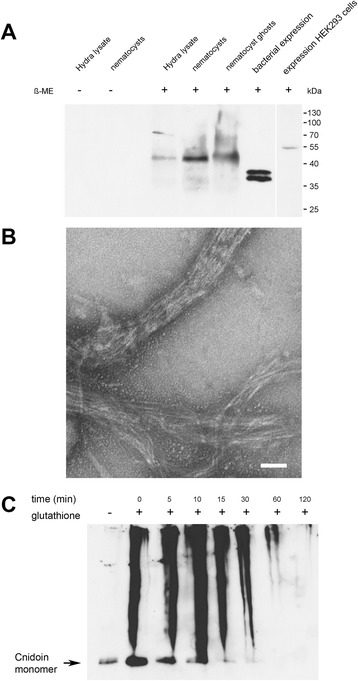
Western blot detection and self-aggregation of Cnidoin. (A) western blot analysis of Cnidoin in isolated nematocysts, the insoluble fraction of nematocysts (ghosts), whole hydra lysate, and after recombinant expression in bacteria and HEK293 cells. (B) transmission electron micrograph of recombinant Cnidoin forming bundles of fibres. Scale bar is 100 nm. (C) western blot analysis of recombinant Cnidoin induced to form disulphide-linked polymers by glutathione treatment.