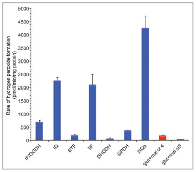
Fig 3.
Hydrogen peroxide generation at different sites in isolated muscle mitochondria. Maximum capacities of sites defined using different combinations of substrates and inhibitors are in blue, actual overall rates during oxidation of glutamate plus malate in the absence of respiratory inhibitors are in red. All rates were either measured in the presence of 1-chloro-2,4-dinitrobenzene to greatly attenuate losses of hydrogen peroxide in the matrix by endogenous glutathione-linked peroxidases,65 or corrected to such measurements using the equations in Quinlan et al.48 and Treberg et al.65 St 4, state 4 (no ATP synthesis); st 3, state 3 (maximum ATP synthesis); OGDH, 2-oxoglutarate dehydrogenase; ETF, electron-transferring flavoprotein and ETF:Q oxidoreductase; DHODH, dihydroorotate dehydrogenase; GPDH, glycerol 3-phosphate dehydrogenase; glut + mal, glutamate plus malate; IF, IQ, IIF, IIIQo, see Table 2. Data are from unpublished observations and references 47, 48 and 59. Values are means ± SEM (n ≥ 3).

