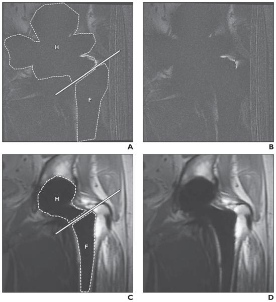
Fig. 1.
45-year-old man with metal-on-metal total hip arthroplasty.
A–D, Comparable coronal 2D fast spin-echo (FSE) (A and B) and coronal multiacquisition variable-resonance image combination selective (MAVRIC SL) (C and D) images. Dashed lines in A and C indicate measured artifact areas in hip (H) and femur (F), and solid lines in A and C indicate basicervical femoral neck line. Artifact areas of hip and femur at 2D FSE (A) are larger than those at MAVRIC SL (C). Bone-metal interface is not visualized on 2D FSE (B) but is well depicted on MAVRIC SL (D). Artifact score was 5 (severe artifact and nonvisualization of bone-metal interface) on 2D FSE (B) and 2 (visible artifacts but well-visualized bone-metal interface) on MAVRIC SL (D) for both reviewers. There is incidental artifact (radiofrequency artifact) on right side of FSE images (A and B).

