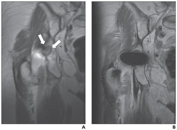
Fig. 4.
73-year-old woman with right metal-on-polyethylene total hip arthroplasty. A and B, Matched coronal proton density–weighted images of multiacquisition variable-resonance image combination selective (MAVRIC SL) (A) and 2D FSE (B) sequences. Well-demarcated, lobulated, intermediate-signal-intensity lesion (arrows, A) is clearly seen in right ischium on MAVRIC SL (A), suggesting osteolysis, but lesion is obscured by metal artifact on 2D FSE image (B). Diagnostic confidence of this image set was 5 (diagnosis cannot be made on 2D FSE but can be confidently made on MAVRIC SL) for both reviewers.

