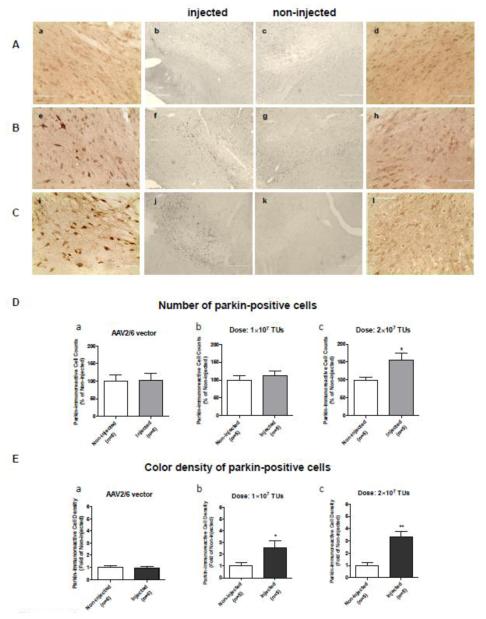
Figure 3.
Validation of parkin overexpression in the substantia nigra pars compacta (SNc) by color immunohistochemistry. Non-coding or parkin-coding adeno-associated virus 2/6 transfer vectors (AAV2/6) were stereotaxically injected in the left SNc of adult male Sprague-Dawley rats with the following coordinates: −5.2 (AP) and −2 (ML) from Bregma, −7.6 (V) mm from the dura. After 3 weeks, the rats were sacrificed by perfusion. (A-C) Representative images of parkin immunolabeling (DAB) in the SNc of rats microinjected with (A) the non-coding AAV2/6 (a-d), (B) 1×10⁷ TUs of AAV2/6-parkin (e-h), and (C) 2×10⁷ TUs of AAV2/6-parkin (i-l), respectively (left: injected and right: non-injected; magnification: a, d, e, h, i, l - 40x; b, c, f, g, j, k - 10x). Injected hemispheres show higher parkin immunostaining than non-injected hemispheres (e, f vs. g, h and i, j vs. k, l), indicating successful dose-dependent overexpression of parkin. Microinjections of the non-coding vector suspensions (1×10⁷ or 2×10⁷ TUs) did not affect endogenous parkin levels (a-d). (D) Quantification of parkin overexpression in the SNc by counting of parkin-positive cells. There was no difference in number of parkin-positive cells between the non-coding AAV2/6-injected and non-injected SNc (p>0.05, unpaired two-tailed Student’s t-test, t=0.1190, df=10, n=6). SNc injected with parkin-coding AAV2/6 (1×10⁷ or 2×10⁷ TUs of AAV2/6-parkin) showed dose-dependent increase in number of parkin-positive cells (+12% and +54%, respectively, as compared to contralateral SNc); however, the 12% increase was not statistically significant (unpaired one-tailed Student’s t-test; 1×10⁷: t=0.6077, df=8, p>0.05; 2×10⁷: t=2.444, df=8, p<0.05; n=5). (E) Quantification of parkin overexpression in the SNc by measurement of color density in parkin-positive cells. Injection of the non-coding AAV2/6 did not affect color density in parkin-positive cells (p>0.05, unpaired two-tailed Student’s t-test, t=0.1965, df=10, n=6). SNc injected with parkin-coding AAV2/6 (1×10⁷ or 2×10⁷ TUs of AAV2/6-parkin) showed dose-dependent increase in color density in parkin-overexpressing cells (medium to light brown) (2.5 fold and 3.3 fold, respectively) as compared to parkin-positive cells contralateral SNc (endogenous parkin: light brown) (unpaired one-tailed Student’s t-test; 1×10⁷: t=2.340, df=8, p<0.05; 2×10⁷: t=4.850, df=8, p<0.01; n=5). *p<0.05, **p<0.01 injected vs. corresponding non-injected SNc. Data are presented as mean ± SEM. Bar in a,d,e,h,i,l: 100 μm; bar in b,c,f,g,j,k: 400 μm. Abbreviations: TUs, transducing units.

