Figure 4.
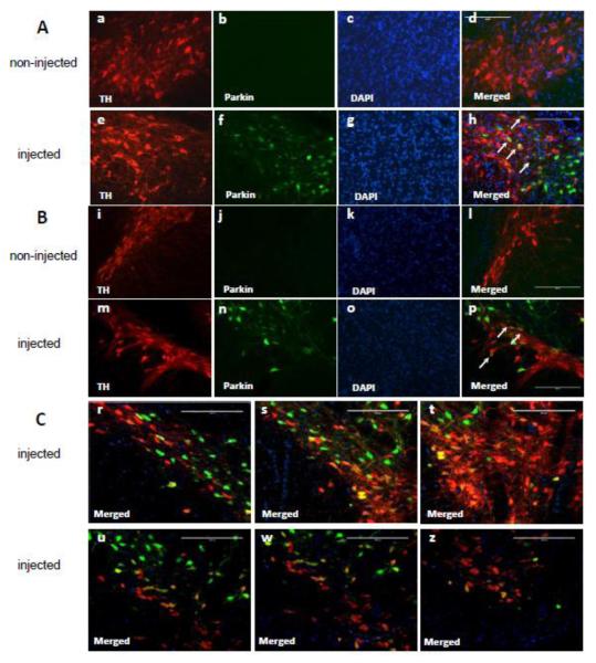
Validation of parkin overexpression in the substantia nigra pars compacta (SNc) by immunofluorescence. Representative images of parkin and tyrosine hydroxylase (TH) immunofluorescent labeling in the SNc: (A) area adjacent to the VTA, (B) middle area of the SNc. In non-injected SNc, parkin immunostaining was not detectable (Ab and Bj). In microinjected SNc, immunolabelling for parkin (green) colocalized with immunolabeling for DAergic TH (red), indicating parkin overexpression in most of the DA neuronal cell bodies (Ah and Bp, white arrows). Parkin-positive neurons were also observed in the ventral tegmental area (Ah). (C) Two coronal planes of the SNc along the rostro-caudal axis. More parkin was overexpressed in DA neurons located closer to the injection site (upper panel) than in DA neurons located away from the site (lower panel). Bars: 200 μm.

