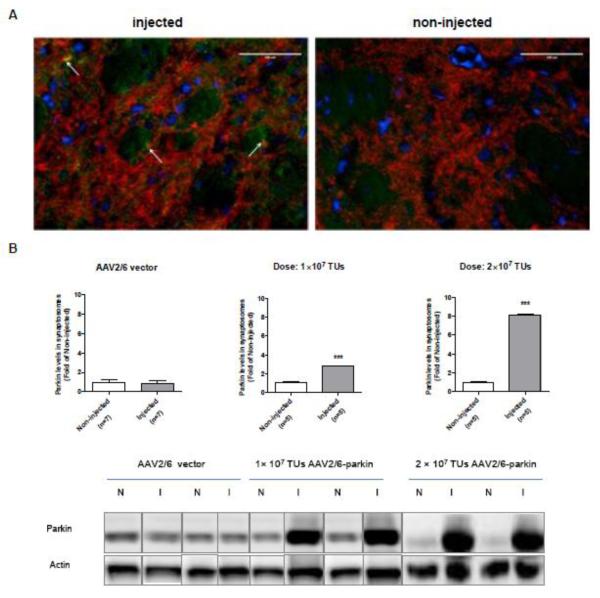
Figure 5.
Validation of parkin overexpression in the striatum. Rats were microinjected with the non-coding AAV2/6 (1×10⁷ or 2×10⁷ TUs) or AAV2/6-parkin (1×10⁷ or 2×10⁷ TUs) into the left SNc, and sacrificed 3 weeks later. (A) Representative image of double immunostaining for parkin and DAergic marker tyrosine hydroxylase (TH) and (B) quantification of parkin levels in striatal synaptosomes by western blotting. (A) Parkin immunostaining is not clearly visible in merged image because of the bright TH staining. Nevertheless, the co-localization of parkin and TH is apparent (A, white arrows). Bars: 100 μm. (B) Representative blots of parkin (52 kDa) and corresponding actin loading control from striatal synaptosomal preparations. Following intranigral microinjection of 1×10⁷ and 2×10⁷ TUs of AAV2/6-parkin, the levels of parkin increased 3 and 8.5 fold, respectively, in ipsilateral striatum as compared to the contralateral striatum (p<0.001, unpaired, one-tailed Student’s t-test; 1×10⁷: t=11.92, df=8; 2×10⁷: t=6.209, df=8; n=5). Microinjections of either dose of the non-coding AAV2/6 resulted in no statistically significant changes in striatal parkin levels (western blot: first 4 bands, 1×10⁷ and 2×10⁷ TUs, respectively) (p>0.05, unpaired two-tailed Student’s t-test, t=0.6062, df=12, n=7). The data are expressed as mean ± SEM. ***p<0.001 injected vs. corresponding non-injected striatum. Abbreviations: AAV vector, adeno-associated viral vector, I, injected; N, non-injected; TUs, transducing units.

