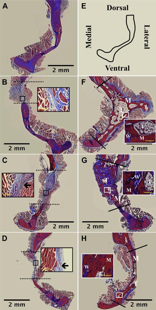
Fig. 7.
Histological images of bone regeneration in section of rat hemi-mandibles 16 weeks post-surgery. Mandibles were sectioned along the coronal plane of the decalcified defect area and stained with Masson's Trichrome. (A) Normal bone and (E) a schematic of the anatomical orientation. Animals administered (B) no treatment, (C) kerateine scaffold, or (D) kerateine gel without rhBMP-2 were unable to bridge the defect (indicated by dashed line). (F) Absorbable collagen sponge, (G) kerateine scaffold, or (H) kerateine gel loaded with 5 μg rhBMP-2 were able to close the defect (solid line) with mature (M), woven (W) and trabecular (T) bone. Each main image is stitched from images collected with a 4× objective; scale bar denotes 2 mm. For B, C, D, F, G, and H insets are images taken with 20× objective (scale bar indicates 0.2 mm). Insets for no treatment (B) and kerateine carriers without rhBMP-2 (C &D) highlight the collagen fiber orientation as well as remaining kerateine carrier after 16 weeks (arrows). Insets for rhBMP-2 treatment groups highlight varying stages of bone formation.

