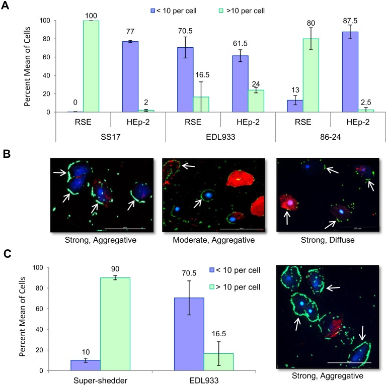
Figure 7. Adherence patterns of O157 strains SS17, EDL933, and 86–24-SmR to RSE cells.
(A) Quantification of SS17, EDL933, and 86–24-SmR adherence to RSE and HEp-2 cells in the presences of D+Mannose. SS17 displays a strong adherence to RSE cells and only moderate adherence to HEp-2 cells. (B) Immunofluorescence stained slides reveled that both SS17 and EDL933 displayed an aggregative adherence pattern on RSE cells while 86–24 displayed a diffuse pattern. All strains demonstrated a diffuse adherence pattern on HEp-2 cells (not shown). (C) Average of adherence levels for nine super-shedder isolates, which all displayed strong, aggregative adherence to RSE cells, that can be seen in the representative immunofluorescence stained slide. Arrows point to adherent bacteria (green). Slides are shown at 40x magnification with RSE cytokeratin (red) and nuclei (blue).

