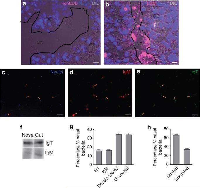
Figure 2. Trout nasal bacteria are coated by secretory immunoglobulins.
(a) Differential interference contrast images of trout olfactory organ stained with NONEUB oligoprobe by fluorescence in situ hybridization. Note that no bacterial staining was observed (N = 5). (b) Differential interference contrast images of trout olfactory organ stained with EUB338 oligoprobe (magenta) that stains ~90% of all eubacteria. Abundant bacteria (magenta) can be observed in the lumen of the olfactory organ and some within the olfactory epithelium (N = 5). Nuclei (blue) are stained with the DNA-intercalating dye DAPI. Scale bar, 20 μm. (c–e) Fluorescent microscopy images of trout nasal bacteria stained with a DAPI-Hoeschst solution (blue; c), anti-IgM (red; d) or anti-IgT (green; e). Orange arrows indicate bacteria that are positive for DAPI, IgM and IgT (double-coated population). Scale bar, 20 μm. (f) Immunoblot analysis of IgTand IgM on nasal-associated bacteria and gut luminal bacteria (as a positive control). (g) Percentage of trout nasal-associated bacteria that are uncoated, coated with IgT, IgM or both IgTand IgM quantified from immunofluorescence microscopy images. (h) Percentage of trout nasal-associated bacteria that are uncoated or coated with at least one antibody isotype quantified from immunofluorescence microscopy images. Data (mean±s.e.) are representative of three independent experiments (N = 6).

