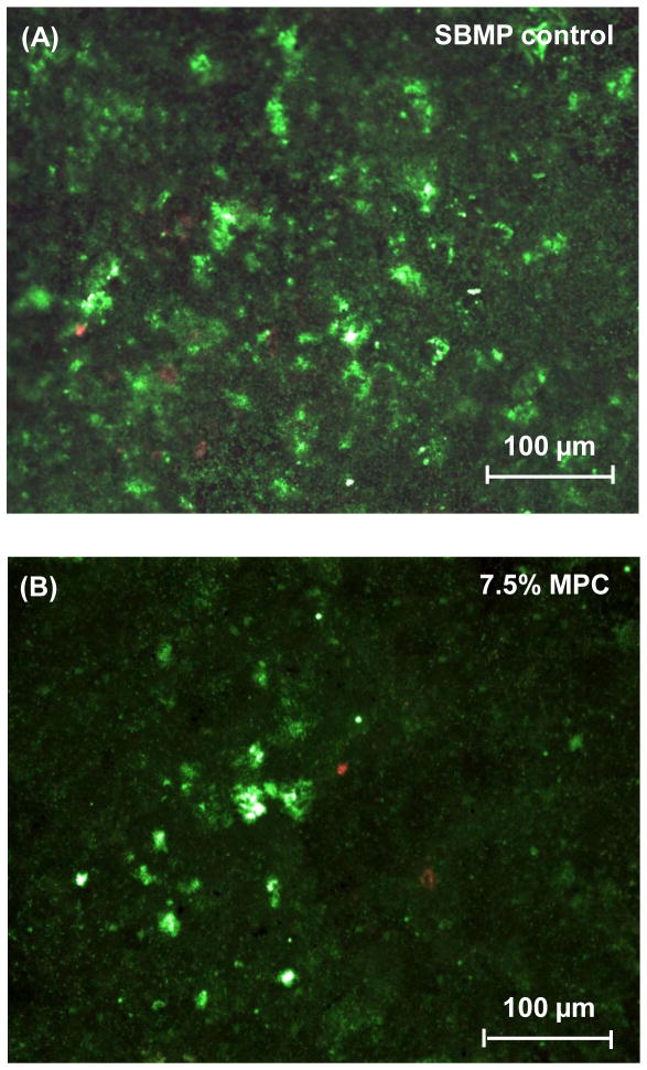
Fig. 3.
Representative live/dead staining images of dental plaque microcosm biofilms grown for 2 days on resin disks: (A) SBMP control, (B) SBMP + 7.5% MPC. In (A), biofilms on control disks had primarily live bacteria covering the entire disk. In (B), substantial decreases in bacterial adhesion occurred when MPC was incorporated into primer and adhesive. The live bacteria were stained green, and the dead bacteria were stained red.

