Figure 1.
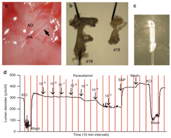
Paracetamol-induced constriction of the ex vivo ductus arteriosus. The ductus arteriosus (arrow) of fetal mice (a) was sharply excised from surrounding tissues. The ductus of term (day 19) and preterm (day 15) fetuses were removed along with a vascular segment extending from the base of the pulmonary artery to the transverse aortic arch (b) (shown with 100 μm dissecting pins). This vascular bloc was mounted on glass pipette tips and secured by tying back the branch pulmonary arteries and the aortic arch, in order to isolate the entire length of the ductus for myography studies (c). Pressure myography was performed to study ductus tone and examine drug-induced changes in lumen diameter (d). After pressurization and equilibration, mounted vessels were briefly exposed to 50 mmol/l KCl to verify contractile potential, then allowed to return to baseline tone. Cumulative exposure to increasing paracetamol demonstrated concentration-dependent constriction of the day 19 ductus (d). Constricted vessels were then exposed to sodium nitroprusside, a potent nitric oxide donor, to document vasodilatory capacity. Terminal exposure to KCl ensured viability of the preparation at the end of each experiment.

