
Figure 1.
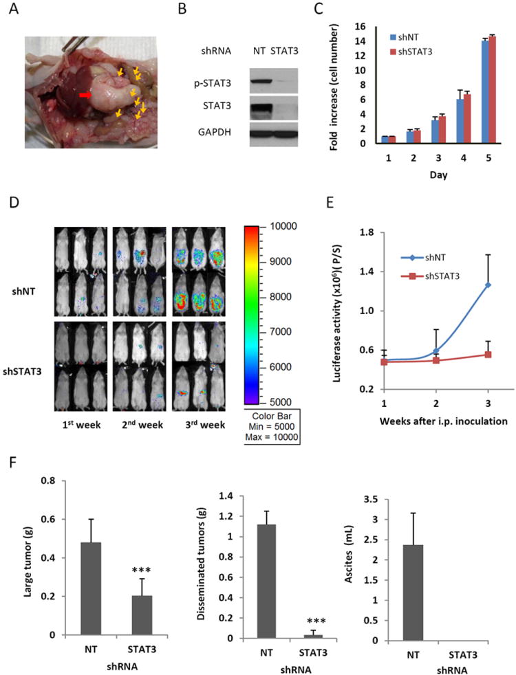
Knocking down STAT3 expression using RNA interference suppresses peritoneal metastasis and ascites production in NSG mice. (A) Representative view of the peritoneal cavity of an athymic nude mouse from 8 mice inoculated with SKOV3-M-Luc cells. Red arrow: large primary tumor; yellow arrows: small tumor nodules. (B) STAT3 expression was blocked in SKOV3-M-Luc cells with shRNA targeted against STAT3. Cells were transduced with STAT3 shRNA or non-targeted (NT) shRNA, and expression of total STAT3 and phosphorylated STAT3 was determined by Western blot analysis. Results are representative of 3 experiments. (C) In vitro proliferation assay. Both shNT cells and shSTAT3 cells were plated and counted each day. (D-E) Luciferase images show cancer progression in mice. STAT3 deficient cells (shSTAT3) and STAT3 active cells (shNT) were inoculated into the peritoneal cavity of NSG mice. Luciferase activities were measured each week after initial cell inoculation (D) and quantified (E). (F) Effect of STAT3 knockdown on tumor burden and ascites volume. At the end of experiment, mice were euthanized. Large primary tumors and small tumor nodules were excised and weighed. Ascites was collected and the volume was measured. n=5-8, ***, P<0.0005, vs. shNT control.