
Figure 1.
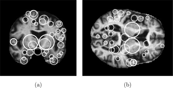
Illustrating scale-invariant features extracted in an MR volume. Circles represent the locations and scales of features in (a) a coronal slice and (b) an axial slice. Solid circles indicate features present in both slices, dashed circles indicate features present in a single slice. Note how feature scales reflect the spatial extent of the underlying anatomical structures, e.g. the width of sulci or ventricles. Here, features are extracted using the DOG operator (Lowe, 2004) and features shown are located within 2 voxels of the corresponding slice through in the 3D volume.