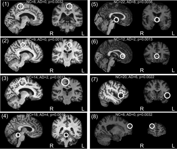
Figure 5.
Examples of the eight most significant NC-related features, shown in sagittal and coronal slices. The feature occurrence frequencies within 75 AD and 75 NC subjects and associated uncorrected p-values are given. Most features lie near the mid-sagittal plane (1-6), and reflect stable un-atrophied cortical (1-3) or sub-cortical (4-6) structure. Feature (7) represents an anatomical pattern reflective of natural age-related atrophy, feature (8) represents a distinctive cortical fold.

