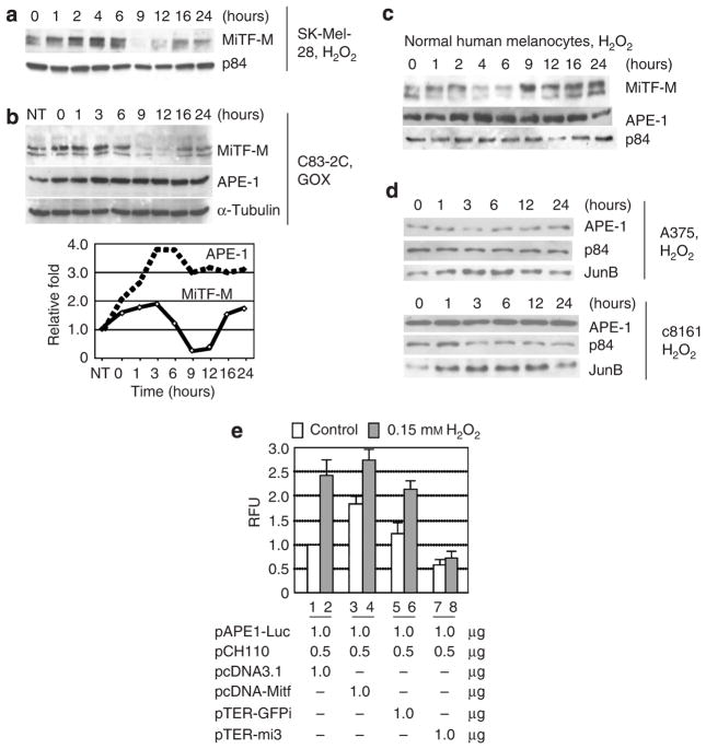
Figure 3. MiTF is phosphorylated and required for APE-1 induction after ROS treatment.
(a) Western blot analysis of MiTF in SK-Mel-28 cells after H2O2 treatment. The Sk-Mel-28 cells were treated with 0.1mM H2O2 and collected for western blot analysis at the indicated time points. p84 serves as a loading control. (b) MiTF and APE-1 accumulation in c83-2C cells after GOX (150mUml−1) treatment. Cells were incubated with GOX for 1 hour, washed with 1 × phosphate-buffered saline, and then returned to incubator with fresh media (time 0). The bottom graph is quantiated MiTF (total) and APE-1 protein levels based on the western blot on top. (c) Western blot analysis of MiTF and APE-1 accumulation in normal human melanocytes after treatment with 0.1mM H2O2. (d) Western blot analysis of APE-1 accumulation in two MiTF-negative melanoma cells A375 (top) and c81-46A (bottom) after 0.15mM H2O2 treatment. (e) Induction of APE-1 promoter activity requires MiTF. SK-Mel-28 cells were transfected with pAPE-Luc and one of the following plasmids: pcDNA3.1, pcDNA-MiTF, pTER-mi3, or pTER-GFPi, together with pCH110. Cells were treated with 0.1mM H2O2 36 hours after transfection and luciferase activity was assayed 48 hours after transfection.

