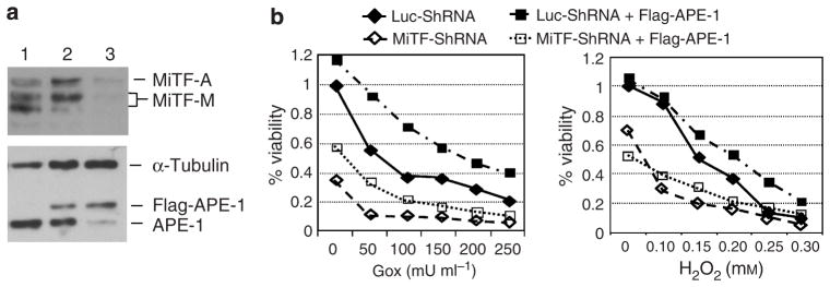
Figure 5. Over-expression of APE-1 rescued ROS-induced cell viability in MiTF-depleted cells.

(a) Concomitant knock-down of MiTF and over-expression of APE-1 in SK-MEL-28 cells. Cells were transiently transfected with pFlag-APE-1, and then infected with Ad-shLuc or Ad-shMiTF 24 hours later. (1) Control SKMel- 28 cells; (2) Cells transfected with pFlag-APE-1 and infected with Ad-shLuc; (3) Cells transfected with pFlag-APE-1 and infected with Ad-shMiTF. α-Tubulin serves as a loading control. (b) Cells with or without MiTF knock-down or APE-1 overexpression were treated with various concentration of glucose oxidase (Gox, left) or H2O2 (right) 48 hours post-infection, and MTT assay was performed to measure cell viability. All data is normalized to cells with Luc-ShRNA transfection.
