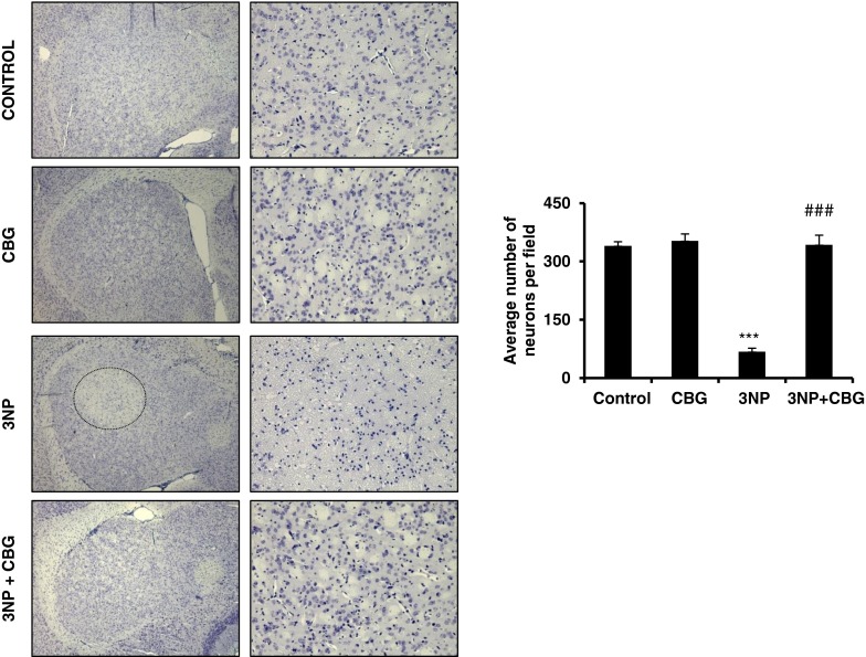
Fig. 2.
Systemic administration of 3-nitropropionic acid (3NP) leads to a progressive and selective degeneration in the striatum. (Left) Cresyl violet staining was performed on brain sections from control, cannabigerol (CBG)-, 3NP-, and 3NP + CBG-treated mice. Low (left column) and high magnification (right column) showing the selective loss of cells in the striatum at day 5 (pale region, outlined). This lesion was not detectable in the group that received CBG. Images were acquired by using light microscopy. (Right) Quantification of Nissl-positive cells in the mouse striatum. Total average number of neurons (100× magnification) is shown. Values are expressed as means ± SEM for 6–8 animals per group. Data were subjected to one-way analysis of variance followed by the Student–Newman–Keuls test. ***P < 0.001 when comparing the control group with the 3NP and CBG group. ### P < 0.001 when comparing the 3NP group with the 3NP + CBG group

