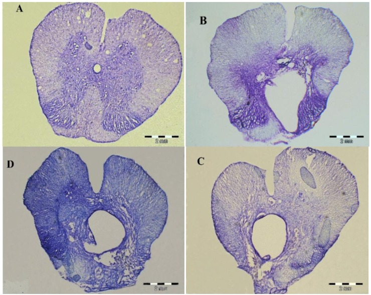
Figure 4.
10 µm thick cross sections of spinal cord segments T8-9 of sham operative group (A), control group (B), hADSCs group (C) and ChABC group (D) at 8 weeks after surgery. Spinal cord tissue was stained with Cresyl violet. A large cavity was shown from non treated animals. The cavity formation was reduced in treated animals

