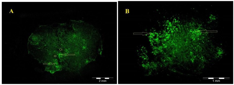
Figure 5.
(A) Low magnification of the GFP-positive hADSCs (green) located within the lesion 8 weeks after transplantation. (B) hADSCs in A are shown at higher magnification. Stars indicates the cystic cavity and arrow indicates GFP-positive hADSCs that migrating into the host tissue around the cystic cavity

