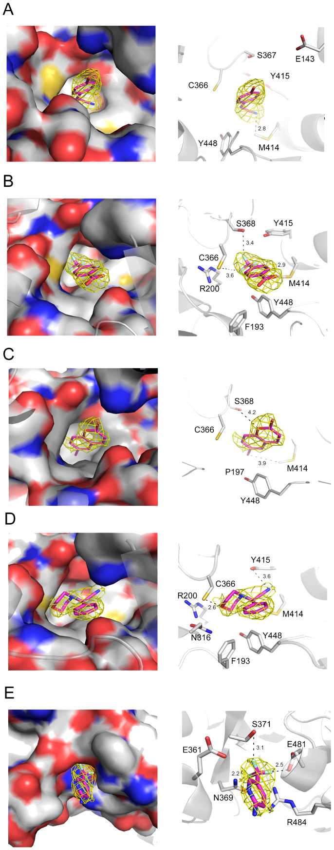
Figure 3. X-ray crystal structures of five fragments found to bind the allosteric site at the palm pocket (A, B, C, D) or at the thumb pocket (E).
Crystal structures of bound fragments 114 (A), 204 (B), 117 (C), 328 (D) and 162 (E) are shown. The Fo–Fc omit electron density maps are shown as a yellow mesh contoured at 3σ around the fragments. Right panels in A, B, C, D and E depict the amino acids found to interact with the fragments, represented by dashed black lines. Carbon atoms are depicted by gray, oxygen by red, nitrogen by blue and sulfur by yellow colors.

