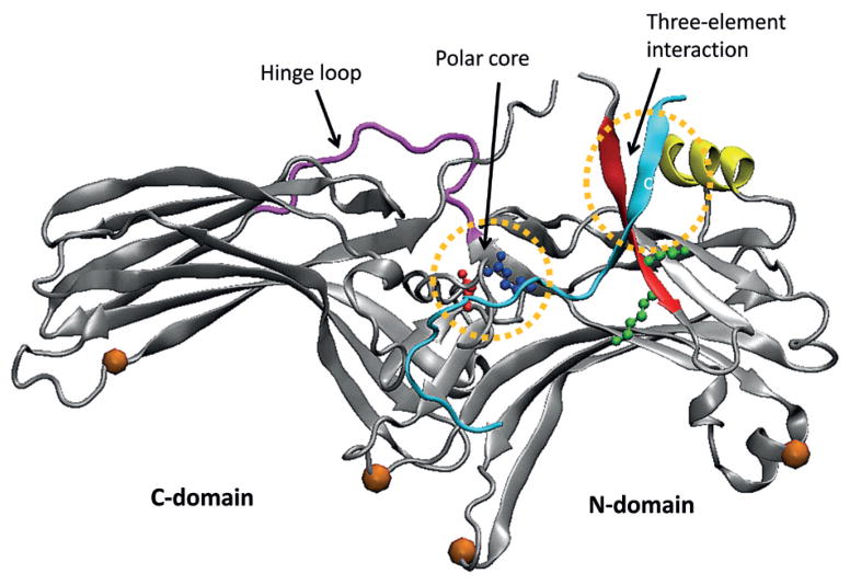
Figure 5. Crystal structure of arrestin (PDB id: 1CF1) with characteristic elements indicated.
Orange balls indicate regions that change upon GPCR binding but are not directly involved in the interaction with receptor. Colored residues are important for arrestin stability (a salt bridge in blue and red in polar core region) or initial recognition of receptor (two Lys residues in green).

