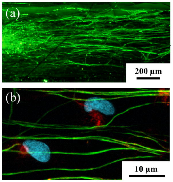
Figure 8.
(a) Confocal image of DRG neurites cultured on the aligned electrospun PEDOT microfibers. Beta-tubulin (green), a structural component of neurons, indicates that the DRG neurites followed the direction of fiber alignment (horizontal). (b) Schwann cells are identified by co-localization of s100 (red) and DAPI (blue). Scale bars: 200 μm (a) and 10 μm (b).

