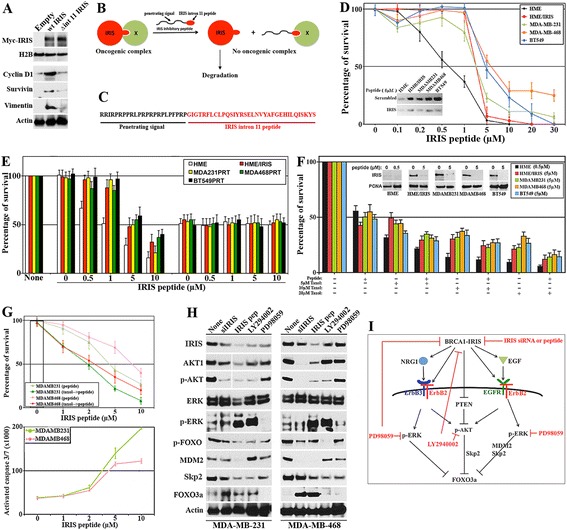
Figure 2.

BRCA1-IRIS inhibitory peptide effect on TNBC cells survival, in vitro . (A) Expression of indicated proteins in HME cells transfected with empty vector, Myc-tagged wild-type BRCA1-IRIS (wt IRIS) or Myc-tagged mutant BRCA1-IRIS (missing the intron 11 domain, Δint 11 IRIS) for 48 h. (B) Schematic representation of the proposed function of BRCA1-IRIS intron 11 domain and peptide. (C) BRCA1-IRIS inhibitory peptide; black sequence is penetrating signal and red sequence is BRCA1-IRIS intron 11 domain. (D) Survival of the indicated cells exposed to increasing concentrations of IRIS peptide. Values are means of triplicates done three separate times. Inset: effect of 5 μM of scrambled or IRIS peptide on BRCA1-IRIS expression in the indicated cell lines. (E) Effect of increasing concentrations of IRIS peptide on the indicated cell lines following luciferase or BRCA1-IRIS silencing. Values are means of triplicates done three separate times. (F) The synergistic effect between IRIS peptide (5 μM) in the indicated TNBC cell lines and 0, 5, 10 and 20 μM of paclitaxel. Values are means of triplicates done three separate times. Inset: effect of 0.5 μM (HME) or 5 μM (other cell lines) of IRIS peptide on BRCA1-IRIS expression. (G, upper) Effect of paclitaxel at 1 μM alone (white bar), increasing concentrations of IRIS peptide alone (light-colored lines) or the combination (dark-colored lines) on the survival of indicated cells. Values are means of triplicates done three separate times. (G, lower) The gradual increase in activated caspase 3/7 in the indicated cells following exposure to increasing concentrations of IRIS peptide. Values are means of triplicates done three separate times. (H) The expression of the indicated proteins in MDA-MB-231 and MDA-MB-468 cells following transfection of BRCA1-IRIS siRNA, or the exposure to 5 μM IRIS peptide, 10 μM of PI3′K/AKT inhibitor (LY294002) or ERK1/2 inhibitor (PD98059). (I) Schematic representation of the data in Figure 2. HME, human mammary epithelial cells; TNBC, triple negative breast cancer.
