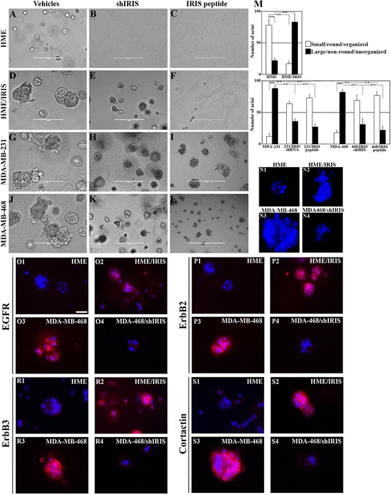
Figure 3.

BRCA1-IRIS overexpression promotes aggressiveness in HME cells, while inactivation inhibits it in TNBC cells. Representative images showing acini formation in low growth factor matrigel-coated wells in the presence of vehicle, shIRIS or IRIS peptide in HME (A-C) HME/IRIS (D-F) MDA-MB-231 (G-I) and MDA-MB-468 (J-L) cell lines at day 10. IRIS peptide was added at 0.5 μM (HME) or 5 μM (the other cell lines). Scale bar in A-L = 400 μm. (M) Quantitative analysis of the number and phenotype of acini formed using HME and HME/IRIS (upper) or MDA-MB-231 and MDA-MB-468 (lower) following the above mentioned treatments at day 10. (N1-N4) Acini formed by HME, HME/IRIS, MDA-MB-468 and MDA-MB-468/shIRIS, respectively. The expression of the indicated proteins in acini formed using HME (O1, P1, R1 and S1), HME/IRIS (O2, P2, R2 and S2), MDA-MB-468 (O3, P3, R3 and S3) and MDA-MB-468/shIRIS (O4, P4, R4 and S4) cells. Scale bar in O1-S4 = 100 μm. HME, human mammary epithelial cells; TNBC, triple negative breast cancer.
