Figure 4.

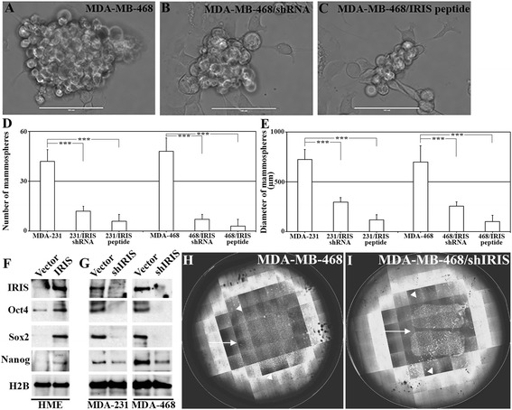
BRCA1-IRIS overexpression promotes tumor-initiating phenotype in HME cells, while inactivation suppresses it in TNBC cells. (A-C) Representative images showing mammosphere formation in MDA-MB-468 cells following control treatments (A), BRCA1-IRIS silencing (B) or inactivation using IRIS peptide (C) at day 10. Scale bar in A-C = 1,000 μm. Quantitative analysis of the number (E) or diameter (F) of mammospheres developed using MDA-MB-231 or MDA-Mb-468 cells after vehicles, BRCA1-IRIS silencing or BRCA1-IRIS inactivation using IRIS peptide. (G) The expression of the indicted stemness biomarkers in HME or BRCA1-IRIS overexpressing HME cells or MDA-MB-231 and MDA-MB468 expressing or silenced from BRCA1-IRIS. Representative images of the migration of MDA-MB-468 (H) or MDA-MB-468 expressing IRIS shRNA (I). In both images arrows show intervening spaces left by the insert that were filled by MDA-MB-468 (24 h later) and not in BRCA1-IRIS-silenced MD-MB-468 and in both images arrowheads show the distance MDA-MB-468 cells travelled outward and the lack of such migration in BRCA1-IRIS-silenced MDA-MB-468 cells. HME, human mammary epithelial cells; TNBC, triple negative breast cancer.
