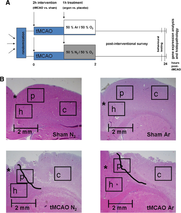
Figure 1.

Study procedure and exemplary histological graphics. A simplified graphic of the study procedure is shown in A. After randomization animals received i.p. anesthesia and tMCAO or sham surgery. 1 h after the occlusion of the MCA with a thread treatment with 50% argon/50% oxygen or placebo (a 50% nitrogen/oxygen-mixture) was performed by mask inhalation for another hour. After that, treatment and MCA occlusion were terminated. Animals were left to recover from the procedure. 24 h later after behavioral testing, animals were sacrificed and brains obtained for further experiments. Example pictures of the HE-stained penumbra regions (slice 3) from the tMCAO + argon (MCAO Ar) and tMCAO + placebo (MCAO N2) groups vs. corresponding regions from sham + argon (Sham Ar) and sham + placebo (Sham N2) are shown in B. The demarcation zone between penumbra and necrotic tissue is tagged with a bold black line. The interhemispheric fissure is asterisked. The positions of the images taken for cell evaluation and quantification are marked by black rectangles (h healthy cortex; p penumbra; c ischemic core).
