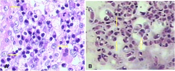
Figure 4.

Histopathology of cervical vertebral (periodic acid-Schiff stain, 400×) (A) and bone marrow samples. Numerous intracellular yeast-like or sausage-like cells measuring 2–3 μm in diameter with a transverse septum were observed (arrows, periodic acid-Schiff staining, 1000×) (B).
