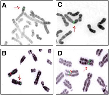
Figure 2.

Evidence on FISH analysis of the Xp11.22 deletions in the patient (A, C) and his mother (B, D) lymphocytes attested by the absence of one red signal on metaphase spread for the BAC clone RP3-519 N18 for the first deletion (control BAC clone RP11-122 F2 in green) and for the BAC clone RP11-637B23 for the second deletion (control BAC clone RP11-258C19 in green).
